Figures
|
|
|
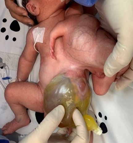
Figure 1
HT with omphalocele. |
|
|
|
|
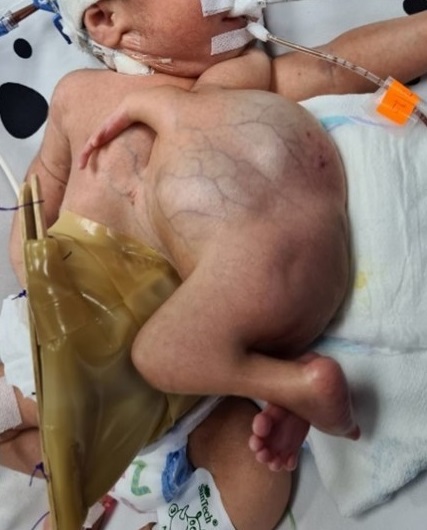
Figure 2
Omphalocele was covered with a temporary silo |
|
|
|
|
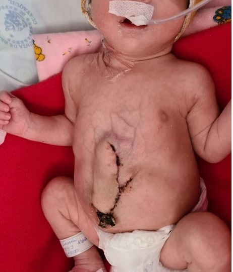
Figure 3
Final outcome after excision of the HT and omphalocele |
|
|
© 2021, Acosta Farina et al
Received Day: 21 Month: 01 Year: 2021 Accepted Day: 07 Month: 03 Year: 2021 J Neonatal Surg. 2021; 10: 16. DOI: 10.47338/jns.v10.938 |
A male baby (weight: 3150g) was born through cesarean section at 35 weeks of gestation to a 32-year-old primigravida with a history of substance abuse. APGAR scores were 7,8 and 8 at the 1st, 5th, and 10th minutes, respectively. The baby developed respiratory distress soon after birth requiring intubation and mechanical ventilation. Physical examination revealed conjoined twins with the autosite having an abdominal wall defect consistent with omphalocele and the parasitic twin joined to the autosite at left hemithorax (Fig. 1). The parasite twin had two upper limbs, two lower limbs, and a phallus with an absent scrotum. An echocardiogram of the autosite showed an interventricular septal defect. A contrast-enhanced abdominal CT scan revealed a parasite twin having an ectopic kidney at the level of epigastrium with associated omphalocele containing a pseudocyst. There was also a large cystic structure suggestive of megabladder. The arterial supply to the parasite was from the internal mammary and superior mesenteric arteries of autosite.
On the first day of life, a temporary silo was placed with a hydrocolloid patch to cover the omphalocele (Fig. 2). After thorough investigations and initial stabilization, surgical excision of the parasite twin was performed on the third day of life. At surgery, the parasite twin was found to have upper and lower limbs supplied by the superior mesenteric and internal mammary arteries of autosite along with the presence of a single kidney, ureter, small and large bowels. The autosite twin had a 10 x 10 cm omphalocele with the liver being its major content. The contents of omphalocele were completely reducible, but primary closure of the abdominal wall was not done to avoid a possible abdominal compartment syndrome. A defect at the anterior portion of the diaphragm was also found which was primarily repaired with a non-absorbable suture. A polypropylene mesh covered with a plastic patch was used to cover the defect created after the excision of the parasitic twin. On the 5th postoperative day, the polypropylene mesh was removed from the abdominal wall, and closure of the abdominal wall defect was done with random rotational flaps and reinforced with a polytetrafluoroethylene patch, which was fixed to the aponeurosis (Fig. 3). On the seventh postoperative day, oral nutrition was started and increased gradually as tolerated. The baby was discharged and followed up in the outpatient setting without any complications.
Parasitic or heteropagus twins (HT) are asymmetrically united twins where the parasite is dependent on autosite for its nutrition and growth.[1] The first case of HT was reported in France in the sixteenth century and until now very little is known about the exact embryology of this pathology.[2] As a result of the low incidence, literature about HT is limited. In Ecuador, only one case report has been published, which serves as a reference on this condition.[3] In our case, the parasite twin had almost normal upper and lower limbs and a phallus without scrotum joined to the left hemithorax of the autosite. The majority of the patients have other associated anomalies.[1], [4], [5]. In our case, the autosite showed omphalocele and interventricular communication. Ultrasound, CT scan, or MRI are used to identify the internal anatomy of the twins especially identifying their blood supply and shared organs.[2], [4] In our case, CT scan showed shared blood supply to the parasite twin that was originating from the superior mesenteric and left internal mammary arteries of the autosite.
Due to the high anatomical variations in the cases of HT, there is no consensus on the best surgical approach.[4] Therefore, it is advised to perform a thorough physical exam and investigations including advanced imaging to plan the best surgical approach that carries the highest survival and life expectancy of the autosite, whose mortality is as high as 31%.[1] The separation of the parasite twin and simultaneous correction of neural tube defects or omphalocele has been described.[3], [6], [7] The defect created after the excision of the parasite can be closed primarily, but at times it is can be so large that it needs other types of closure using flaps, tissue expanders, and multiple surgeries for a better outcome.[4], [5] In our case defect was large and it was closed by random rotational flaps with a good outcome.
|
|
|
|
Figure 1
HT with omphalocele. |
|
|
|
|
|
Figure 2
Omphalocele was covered with a temporary silo |
|
|
|
|
|
Figure 3
Final outcome after excision of the HT and omphalocele |
|
n1Conflicts of interest. None
n2Source of Support: Nil
n3Author contributions: Author(s) declared to fulfill authorship criteria as devised by ICMJE and approved the final version. Authorship declaration form, submitted by the author(s), is available with the editorial office.
n4Consent to Publication: Author(s) declared taking informed written consent for the publication of clinical photographs/material (if any used), from the legal guardian of the patient with an understanding that every effort will be made to conceal the identity of the patient, however it cannot be guaranteed.
None
| 1. | Ángel Fernández JA, Marín GM, Tocuyo YE. Heteropagus twin. Revista Repertorio de Medicina y Cirugía. 2019; 28:132-6. [Article in Spanish]. |
| 2. | Bishop L, Jones B, Kelley D, Martin C. External parasitic twins. J Pediatr Surg Case Rep. 2019; 40:62–8. |
| 3. | Navarrete Castillo R, Leiva Flores J, Ramírez Rivera J. Heteropagus epigastric twins. About a case. Revista Médica de Nuestros Hospitales. 2013; 19:223-5. [Article in Spanish]. |
| 4. | Dejene, B, Negash SA, Mammo TN, Tadesse A, Getachew H, Derbew M. Heteropagus (parasitic) twins. J Pediatr Surg Case Rep. 2018; 37:44-9. |
| 5. | Sharma G, Mobin SSN, Lypka M, Urata M. Heteropagus (parasitic) twins: a review. J Pediatr Surg. 2010; 45:2454–63. |
| 6. | Malik M, Bahadur Singh U, Hedge S, Mahajan JK, Samujh R. Omphalocele and epigastric heteropagus: implications and treatment. Oxf Med Case Rep. 2018; 10:342–5. |
| 7. | Ahmed K, Mahdi BD, Hayet Z, Hamdi L, Mohamed J, Riadh M. Thoracic heteropagus conjoined twins associated to an omphalocele: Report of a case and complete review of the literature. Afr J Paediatr Surg. 2016; 13:209-12. |