Figures
|
|
|
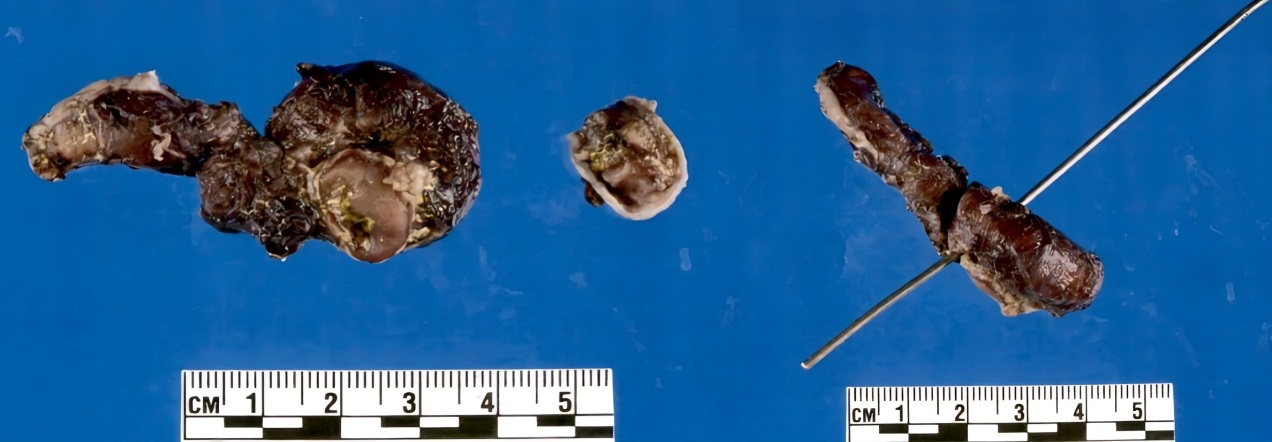
Figure 1
Pathologic specimen showing the perforated Meckel’s diverticulum that was surgically removed from our patient. |
|
|
© 2023, Koura et al.
Received Day: 17 Month: 08 Year: 2022 Accepted Day: 31 Month: 10 Year: 2022 J Neonatal Surg. 2023; 12: 7. DOI: 10.47338/jns.v12.1127 |
|
Keywords: Meckel’s diverticulum, Perforation, Small bowel obstruction. |
||
Meckel’s diverticulum is a congenital outpouching in the small intestine that develops from incomplete obliteration of the embryologic vitelline duct. Meckel’s diverticulum occurs in 2% of the population and most symptomatic cases present in children aged 2 years with lower gastrointestinal bleeding.[1] Spontaneous perforation is an uncommon complication of Meckel’s diverticulum occurring in less than 10% of patients, usually associated with heterotopic mucosa or diverticulitis. [2]
Only a few cases of spontaneous perforation of Meckel’s diverticulum are published in the literature with little data describing the cause of perforation. We present a case of perforated Meckel’s diverticulum in a preterm infant that presented with abdominal distension and partial small bowel obstruction.
A 22-year-old primary gravid delivered a 495-gram male, at 26 weeks and 5 days gestational age, by cesarean section due to persistent reversal of the end-diastolic flow of the umbilical artery. The pregnancy was complicated by intrauterine growth restriction and concern for hydrops fetalis with pericardial effusion and ascites. The APGAR scores were 6 and 8 at 1 and 5 minutes, respectively, and resuscitation consisted of oxygen, continuous positive airway pressure (CPAP), and intubation. Throughout his stay in the neonatal intensive care unit (NICU), our team intubated and mechanically ventilated the patient and slowly weaned him to noninvasive mechanical ventilation (NIMV), CPAP, and high-flow nasal cannula (HFNC).
At 5 weeks of life, he developed necrotizing enterocolitis (NEC) that resolved with antibiotic therapy and bowel rest. At 15 weeks of life, he developed an episode of firm abdominal distension without changes in bowel movements or emesis. An abdominal radiograph demonstrated dilated loops of the small bowel concerning a small bowel obstruction, therefore we started him on bowel rest and placed an oral gastric tube with the resolution of distension. At 17 weeks of life, he developed a recurrent episode of abdominal distension with a tympanic and tender abdomen, however, continued to have regular bowel movements and no emesis. An abdominal radiograph demonstrated dilated loops of the bowel with decompressed distal bowel, and our team again started him on bowel rest and placed an oral gastric tube. A contrast enema demonstrated no definite strictures present in the colon and an upper gastrointestinal study with small bowel follow-through demonstrated the slow passage of contrast with dilated loops of the small bowel. Serial radiographs demonstrated persistent dilated loops of the small bowel with poor antegrade progression of contrast. At this time, we suspected that the patient had developed a partial small bowel obstruction secondary to a stricture from his prior episodes of NEC.
An exploratory laparotomy showed a perforated Meckel’s diverticulum causing partial small bowel obstruction due to intestinal adhesions. Due to the adhesions, we resected the portion of the small bowel proximal and distal to the perforated Meckel’s diverticulum and repaired it with ileo-ileal anastomosis. Pathology confirmed a Meckel’s diverticulum (5.6 cm x 1.2 cm) with a transmural defect (0.4 x 0.3 cm) with serosal adhesions (Fig. 1) without any ectopic gastric or pancreatic tissue.
Postoperatively, we continued an oral gastric tube for 8 days. He began having bowel function and tolerating feedings by the 10th postoperative day. He remained in the NICU to wean oxygen requirements and ensure tolerance of oral feedings and we discharged him from the hospital at 22 weeks of life.
Common clinical presentations of Meckel’s diverticulum include intestinal hemorrhage, intussusception, and volvulus. [1] Our patient’s perforation led to the development of a partial small bowel obstruction which poses additional risks including intra-abdominal abscess, sepsis, and bowel necrosis if not treated promptly. [3] One study reported a case in which a patient developed rapidly progressive septicemia with E.coli due to perforation of Meckel’s diverticulum. [4]
Initial evaluation of our patient suggested the bowel obstruction developed from a stricture due to the patient’s prior episode of NEC that occurred 15 weeks earlier. However, strictures as a complication of NEC typically occur within 5-7 weeks. [5] Most documented cases of perforation of Meckel’s diverticulum present with abdominal distension, likely from pneumoperitoneum. [4], [6], [7], [8]
Spontaneous perforation of Meckel’s diverticulum has been hypothesized to be associated with heterotopic gastric or pancreatic tissue or diverticulitis, however, in a recent systematic review, 78% of cases were not associated with heterotopic mucosa. [1], [9] The pathology from our patient did not have heterotopic tissue, nor any signs of diverticulitis, suggesting an alternative cause for perforation. It is possible that a perforation of Meckel’s diverticulum occurred during the initial episode of NEC.
In a recent study, it was reported that approximately 85% of cases of perforated Meckel’s diverticulum presented within the first week of life. [1] One report of Meckel’s diverticulum was thought to have perforation due to intestinal immaturity within the first 24 hours of life in a premature infant. [6] This is a less likely explanation for our patient given that the first symptoms of perforated Meckel’s diverticulum did not occur until 15 weeks of life, allowing the gut to mature in our patient.
A recent review reported 22% of cases of spontaneous perforated Meckel’s diverticulum had received oxygen supplementation. [1] McKelvie et al. examined 7 reported cases of perforated Meckel’s diverticulum in neonates ranging from 1-10 days of life and suggested an association with complicated pregnancy or neonatal respiratory distress syndrome (NRDS) shortly after delivery with no identified respiratory pathology. [7] While our patient had respiratory distress at delivery, these prior case reports developed perforation of Meckel’s diverticulum within 10 days of their NRDS, whereas our patient’s developed at 15 weeks of life. However, our patient was placed on multiple ventilation methods including intubation, NIMV, CPAP, and HFNC throughout their NICU stay. Wang suggests mechanical ventilation contributes to the progression of abdominal distension and pneumoperitoneum seen with perforation of Meckel’s diverticulum. [6] Mechanical ventilation administered through nasal prongs and masks has been linked with 30 times increased likelihood of upper and lower gastrointestinal perforation. [10] It is possible that increased pressure in the gastrointestinal tract as a result of chronic mechanical ventilation requirements contributed to the perforation in the patient presented in this case.
Following resection of the perforated Meckel’s diverticulum, postoperative recovery appears to be uncomplicated, with oral feedings initiated as early as the 5th day of surgery. [4] The patient in this case report has done well without postoperative complications.
In conclusion, although Meckel’s diverticulum typically presents in early childhood with intestinal hemorrhage or intussusception, this case report demonstrates that in the neonatal period, it may present as a small bowel obstruction.
|
|
|
|
Figure 1
Pathologic specimen showing the perforated Meckel’s diverticulum that was surgically removed from our patient. |
|
n1Conflicts of interest. None.
n2Source of Support: Nil
n3Author contributions: Author(s) declared to fulfill authorship criteria as devised by ICMJE and approved the final version. Authorship declaration form, submitted by the author(s), is available with the editorial office.
n4Consent to Publication: Author(s) declared taking informed written consent for the publication of clinical photographs/material (if any used), from the legal guardian of the patient with an understanding that every effort will be made to conceal the identity of the patient, however it cannot be guaranteed.
None
| 1. | Liaqat N, Mahomed A, Nayyar S, Akhtar N, Ali S, Haider N. Perforated Meckel’s diverticulum in neonates: a report of six cases and systematic review of the literature. Ann Pediatr Surg. 2022; 18:1-9. |
| 2. | Aguayo P, Fraser JD, St. Peter SD, Ostlie DJ. Perforated Meckel's diverticulum in a micro premature infant and review of the literature. Pediatr Surg Int. 2009; 25:539-41. |
| 3. | Reddy SRR, Cappell MS. A systematic review of the clinical presentation, diagnosis, and treatment of small bowel obstruction. Current Gastroenterol Rep. 2017; 19:28. |
| 4. | Zahraa J, Abu-Ekteish F, Al Bassam A-R, Nosir AA. Perforated Meckel’s diverticulum in a neonate mimicking necrotizing enterocolitis. Pediatr Emer Care. 2003; 19:418-9. |
| 5. | Federici S, De Biagi L. Long-term outcome of infants with NEC. Curr Pediatr Rev. 2019; 15:111-4. |
| 6. | Wang Y-jiao, Wang T, Xia S-lin, Zhang Y-cheng, Chen W-bing, Li B. Perforation of Meckel's diverticulum in a very low birth weight neonate with severe pneumoperitoneum and review of the literature. Turkish J Pediatr. 2019; 61:460. |
| 7. | McKelvie M, Soares-Oliveira M, Wang-Koh Y, Trayers C, Aslam A. Beware the innocent presentation of a spontaneous perforated Meckel diverticulum. Pediatric Emergency Care. 2019;35(12):881-883. |
| 8. | Chang Y, Lin J, Huang Y. Spontaneous perforation of Meckel’s diverticulum without peritonitis in a newborn: report of a case. Surg Today. 2006; 37:1114-7. |
| 9. | Oyachi N, Takano K, Hasuda N, Arai H, Koshizuka K, Matsumoto M. Perforation of Meckel's diverticulum manifesting as aseptic peritonitis in a neonate: Report of a case. Surg Today. 2007; 37:881-3. |
| 10. | Donahue L. Spontaneous intestinal perforation. Neonatal Network. 2007; 26:335-51. |